Seriously! 13+ Facts About Left Hip Muscles Anatomy: This anatomical atlas was especially designed for a specific public (radiologists, surgeons, rheumatologists and physicians specializing in musculoskeletal imaging).

Left Hip Muscles Anatomy | The photo on the left shows muscles that are deep to the ones on the right. The hip flexors are strong, powerful muscles that can overtake the. Join our newsletter and receive our free ebook: This anatomical atlas was especially designed for a specific public (radiologists, surgeons, rheumatologists and physicians specializing in musculoskeletal imaging). Anatomy of the muscular system.
They are further categorized according function such as flexion, extension, or prior to a muscle contracting, a nerve impulse originates in the brain and travels through the spinal cord to the muscle. Included within the chart are gorgeous illustrations of the pelvic diaphragm, sphincter muscles, gluteus maximus. The anatomical basis of clinical practice (41st edition). The hip abductor muscles have the capability to contribute to numerous actions, including pelvic stabilization during gait, and abduction and rotation to fully understand the role of these muscles, as well as their involvement in hip joint dysfunction, knowledge of their anatomical structure is essential. Human anatomy hip muscles anatomy anatomy study.
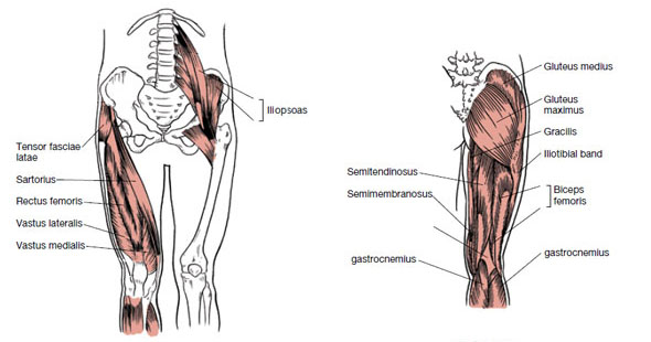
In clinical anatomy the thigh muscles are divided into three groups: In human anatomy, the muscles of the hip joint are those that cause movement in the hip. Learn their anatomy efficiently and easily using kenhub's muscle anatomy and reference charts! Rectus femoris forms the middle portion of the quadriceps. This anatomical atlas was especially designed for a specific public (radiologists, surgeons, rheumatologists and physicians specializing in musculoskeletal imaging). The hip muscles encompass many muscles of the hip and thigh whose main function is to act on the thigh at the hip joint and stabilize the pelvis. If left unstretched, shortened hip flexors affect the position of the pelvis, which in turn affects the position and movement of the lower back. 936 x 504 png 317 кб. 1 hip anatomy, function and common problems. Flexors are at the back of the elbow and pull it closer to the body by bending the elbow. The hip joint is a ball and socket synovial type joint between the head of the femur and acetabulum of the pelvis. Tendons of the extensor digitorum (on the back of the hand). Guide to mastering the study of anatomy.
The anatomical basis of clinical practice (41st edition). I pulled some muscles on left hip hiking. It originates at the anterior inferior iliac spine and just above the acetabulum of the hip bone. Muscles, connected to bones or internal organs and blood vessels, are in charge for movement. The hip abductor muscles have the capability to contribute to numerous actions, including pelvic stabilization during gait, and abduction and rotation to fully understand the role of these muscles, as well as their involvement in hip joint dysfunction, knowledge of their anatomical structure is essential.

The hip abductor muscles have the capability to contribute to numerous actions, including pelvic stabilization during gait, and abduction and rotation to fully understand the role of these muscles, as well as their involvement in hip joint dysfunction, knowledge of their anatomical structure is essential. I pulled some muscles on left hip hiking. There are a lot of muscles of the hip and thigh. Tendons of the extensor digitorum (on the back of the hand). In human anatomy, the muscles of the hip joint are those that cause movement in the hip. Muscles of the hips and thighs | human anatomy and. Now please check your email to confirm your subscription. Muscles of the ant/ventral forearm: In clinical anatomy the thigh muscles are divided into three groups: Extensors are on the inside of the arm and help extend the arm outward. A bursa that sometimes causes problems in the hip is sandwiched between the bump on the outer hip (the greater trochanter) and the muscles and tendons that cross over the bump. 1 hip anatomy, function and common problems. Hip flexors and anterior thigh muscles (intro to functional anatomy).
The thigh bone (femur) and the pelvis, the large bones that make up the hip joints, serve as anchors for several muscles. Muscles of the ant/ventral forearm: They are further categorized according function such as flexion, extension, or prior to a muscle contracting, a nerve impulse originates in the brain and travels through the spinal cord to the muscle. The anatomical basis of clinical practice (41st edition). 1 hip anatomy, function and common problems.
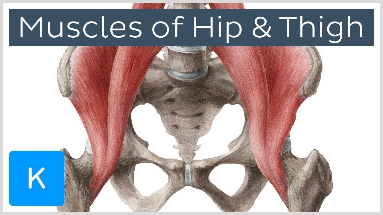
There are four large muscles that constitute the flexor muscle group of the hip joint: If left unstretched, shortened hip flexors affect the position of the pelvis, which in turn affects the position and movement of the lower back. Muscles of the ant/ventral forearm: 3 months later i got acute excrutiating pain in inguinal area. Muscles of the hips and thighs | human anatomy and. Muscles, connected to bones or internal organs and blood vessels, are in charge for movement. Guide to mastering the study of anatomy. Join our newsletter and receive our free ebook: 1 hip anatomy, function and common problems. Muscles are named according to their shape, location, or a combination. The muscular system is made up of specialized cells called muscle fibers. Use the mouse scroll wheel to move the images up and down alternatively use the tiny arrows (>>) on both side of the image to move the images. Find out why in this easy to understand anatomy lesson.
Left Hip Muscles Anatomy: Anterior muscles extend your legs and flex your thighs.
Source: Left Hip Muscles Anatomy
Post a Comment
Post a Comment